微塑料的人体富集及毒性机制研究进展
摘要: 微塑料(MPs)作为一种新型环境污染物已成为当下的研究热点, 有关微塑料的人体健康风险和危害效应机制研究受到了广泛关注.微塑料不断地从环境中迁移并在人体内积累, 其对人群暴露的3个主要途径为经口摄入、呼吸吸入和皮肤接触, 主要暴露介质为食品、饮用水、灰尘和个人护理品.目前已在人体消化系统、呼吸系统、心血管系统和生殖系统的器官、体液及排泄物中检出微塑料, 丰度范围为0~1 206.94 n·g-1.现有的检测分析技术具有不同的适用范围、优势和不足, 针对实验过程中可能污染样品的问题列举了实验室质量保证和质量控制的操作方法.基于动物实验、人体细胞和器官模型的研究阐述了微塑料对人体5大系统造成的潜在健康影响和作用机制, 进入人体后, 微塑料可能通过诱发细胞毒性、线粒体毒性、DNA损伤和细胞膜损伤等效应过程, 进而在人体各系统中引发器官的局部炎症、菌群失调和代谢紊乱等严重后果, 来危害各系统及相关器官的正常功能.最后, 提出了现有研究中普遍存在的不足, 可为未来微/纳米塑料对人体健康影响的研究提供方向.
BAO Ya-bo1,2,WANG Cheng-chen1,2,PENG Wu-guang1,2,NONG Dai-qian1,2,XIANG Ping1,2
Abstract: The effect of microplastics on the ecological environment and human health has become a topical issue, and research on the risks and harmful effects of MPs on human health in particular has attracted widespread attention. Due to the characteristics of small size, low degradability, and easy migration, MPs continuously migrate from the environment to the human body, and their main exposure pathways are oral ingestion, inhalation, and dermal contact, with the main exposure media being food, drinking water, dust, personal care products, etc. MPs have been detected in organs, fluids, and excreta of digestive, respiratory, cardiovascular, reproductive systems, etc. The abundance range of MPs in the human body is 0-1 206.94 particles per gram. After entering the human body, MPs can cause cytotoxicity, mitochondrial toxicity, DNA damage, cell membrane damage, and other effects on human cells and organs, leading to serious consequences such as local inflammation, ecological imbalance, metabolic disorders, etc., in various systems. Owing to their small specific surface area, they can also adsorb pollutants such as heavy metals, organic pollutants, antibiotics, pathogens, and harmful microorganisms, causing combined toxicity and immunotoxicity. In the end, we highlighted general deficiencies in existing studies and provided directions for future research on the influence of MPs on human health.
Key words:microplastics(MPs) contaminants exposure pathways human accumulation toxic effects
微塑料(microplastics, MPs)指直径 < 5 mm的塑料微粒, 是近年来受国内外广泛关注的一种新型污染物.自20世纪50年代大规模生产以来, 塑料被广泛应用于建筑、包装、运输、个人护理产品和医药, 全球塑料产量在1950~2020年间由150 t增长到3.67亿t, 其中2020年我国塑料产量高达1.05亿t[1].据统计, 2019年全球塑料制品回收利用率仅9%, 按传统模式处理率69%(包括:填埋和焚烧), 无人管理而滞留环境的部分占22%[2].塑料制品的滥用和不合理处置是MPs产生的主要原因[3].MPs根据成因可以分为原生微塑料和次生微塑料.原生微塑料指特地生产用于添加到个人护理用品和化妆品中的塑料微粒[4].次生微塑料指环境中原先较大的塑料制品在UV辐射、生物作用和磨损等作用下降解而形成的塑料微粒[5].随着研究发展的需要, 将微塑料依据直径大小进一步细分为:大塑料颗粒(> 25 mm)、中塑料颗粒(5~25 mm)、微塑料颗粒(1 µm~5 mm)、亚微塑料颗粒(100 nm~1 µm)和纳米塑料颗粒(< 100 nm)[6].
由于粒径小、易迁移和降解等特性[7], 微/纳米塑料(M/NPs)广泛分布于全球海洋、淡水、土地和大气生态系统中.近年来, 伊朗新鲜积雪[8]、青藏高原河流[9]、阿尔卑斯山冰川[10]、北极中央盆地地下水[11]和中国海底沉积物[12]中均检测出M/NPs, 其潜在的生态风险受到了广泛关注.M/NPs进入生态系统后, 不仅会影响其生态系统服务功能, 同时对动物、植物和微生物产生有害影响[13].近年来, 越来越多的研究发现M/NPs可以通过口摄入和呼吸吸入进入人体, 由于M/NPs粒径小且比表面积大, 能够作为载体吸附其他有毒污染物(如:重金属、持久有机污染物、抗生素、多环芳烃和多氯联苯等)及有害病原体(如:致病细菌和真菌等)并将它们带入人体内, 从而产生健康危害[3].此外, M/NPs在人体内降解后, 有毒添加剂(如:增塑剂、合成抗氧化剂、阻燃剂、光稳定剂和染色剂等)随之浸出[14, 15], 无疑会加重其毒性.目前, 有关M/NPs的人群暴露及其健康危害机制的研究成为了当下的研究热点.鉴于此, 本文总结了M/NPs的人群暴露途径, 在不同组织器官的富集特征, 及暴露后对人体产生的健康效应机制, 并提出了该领域现有研究的不足和未来可能的热点方向.
1 微/纳米塑料的人体暴露途径
人体与外界环境时刻进行着物质交换(图 1), 摄入食物、呼吸空气和皮肤接触的过程伴随着M/NPs暴露.有研究表明, 经口摄入和呼吸吸入是M/NPs人体暴露主要的途径, 虽然目前缺乏M/NPs穿透皮肤屏障被吸收的直接证据, 但理论上直径 < 100 nm的纳米塑料(NPs)能够穿透皮肤屏障被吸收[16, 17].
1.1 伴随食物经口摄入
研究人员在海产品、饮品、食盐、一次性塑料餐具和果蔬中检测到了M/NPs, 并指出经口摄入是M/NPs最主要的暴露途径.有研究表明, M/NPs能够沿着食物链向更高营养级转移, 最终随着食物转移到人的消化系统中.Nor等[18]建立了儿童和成人终生暴露模型, 估算出一个儿童或成人每天摄入M/NPs的数量的中位数分别为553个与883个.Cox等[19]汇编了来自盐、海鲜、蜂蜜、饮用水和糖的M/NPs污染数据, 并结合饮食习惯估算出美国民众每人每年对M/NPs的摄入量约3.9×104~5.2×104个.近年来, 食物中M/NPs的污染成为人们关注的焦点.
1.1.1 海产品M/NPs污染
已有研究证实了一些浮游动物和滤食性动物能够直接摄入M/NPs.目前, 全球已累计在120多种具有重要渔业价值的物种中发现了M/NPs污染, 其中M/NPs在贝类、鱼类、甲壳类和海藻等海产品中被大量检出[20~22].据统计, 消费者每食用100 g贻贝将摄入70种M/NPs.此外, 海洋生物消化道和鳃中M/NPs的富集更为丰富, 食用完整的或不完全去除内脏的海产品会增加M/NPs对人体的暴露量[23, 24].最近的一项来自韩国的研究也证实, 其民众每人每年通过食用4种常售双壳贝类造成的M/NPs摄入量达212个[25].
1.1.2 饮用水和饮料M/NPs污染
M/NPs普遍存在于各类饮用水和饮料中.其中, 水源污染、处理过程中污染和自来水管管壁脱落是自来水被M/NPs污染的主要原因[26].Kosuth等[27]研究发现自来水中M/NPs检出率高达81%, 平均丰度为5.45 n·L-1, 每人每年因自来水摄入量约为5 800个.因具有易携带性、选择多样性和食品安全性等优点, 瓶装水和饮料的消费日益增长, 自1940~2015年, 法国民众每人每年对瓶装水的消费量从6 L增长至140 L[28].瓶装水和饮料中M/NPs的主要来源是塑料瓶盖与包装瓶.Kankanige等[29]对泰国市场常售的一次性塑料瓶装水进行分析, 结果表明M/NPs平均丰度为(140±19)n·L-1, 粒径约6.5~20 µm, 其主要聚合物类型为聚乙烯(PE)、聚对苯二甲酸乙二醇酯(PET)、聚丙烯(PP)和聚酰胺(PA).相比自来水, 饮用瓶装水导致更高的M/NPs摄入, 若人群仅通过饮用瓶装水而达到推荐饮水量, 则每人每年因此产生的M/NPs额外摄入量将高达9×104个, 若仅饮用自来水, 这一数值将减少到4×10³个[19].可见饮用水M/NPs污染已成为人类摄入M/NPs的重要来源之一, 建议减少饮用塑料包装的水或饮料以有效降低M/NPs的摄入量和健康风险.
1.1.3 食盐M/NPs污染
食盐能提供人体必需元素, 是人类长期的必需品, 然而被M/NPs污染的食盐是一个慢性暴露源.商品盐(包括:海盐、井盐、岩盐和湖盐等)被M/NPs污染的问题已在全球各国广泛报告[30~32].据Lee等[33]研究证实, 全球94%的盐产品中含有M/NPs, 包括27种聚合物类型, 其中PET、PP和PE占绝大多数.食盐被M/NPs污染是多源的, 在采集、风干、加工和运输等过程中, 食盐都可能被M/NPs污染.更有研究发现, 海盐样本中M/NPs的丰度远高于岩盐及湖盐样本[30], 这可能与食盐产生的天然环境中M/NPs污染程度有关.
1.1.4 水果蔬菜的M/NPs污染
2022年, Conti等[34]首次在食用水果(苹果和梨)与蔬菜(胡萝卜、花椰菜、生菜和土豆)中发现M/NPs.Dong等[35]研究发现, 1 µm的聚苯乙烯微塑料(PS-MPs)能够进入胡萝卜根并积累在细胞间隙中, 而0.2 µm的PS-MPs则能够迁移到叶子上, 并且土壤-植物系统中M/NPs的迁移受到重金属的影响.As(Ⅲ)能使得细胞壁变形以及PS-MPs携带负电荷的面积增加, 从而使更大直径的PS-MPs得以进入胡萝卜细胞.此外, Liu等[36]还在食用生鸡蛋中检测出M/NPs, 蛋黄中M/NPs的平均数量高于蛋白, 蒸熟操作后的结果不变.鸡蛋中微塑料可能来自鸡体内, 或是包装和运输过程[37].
1.1.5 塑料包装、餐具和厨具的M/NPs污染
食品接触到塑料包装、一次性餐具和厨具而引发M/NPs污染的问题广泛存在.Du等[38]对中国5个城市常用的外卖餐盒进行研究, 发现所有餐盒中都存在M/NPs, 它们主要源自塑料餐盒内壁脱落和灰尘沉降.高温、机械应力和长时间存放食品都能导致塑料餐具产生更多M/NPs.假设每人每日消耗4~5个一次性塑料水杯, 每人每年将因此摄入3.7×10⁴~8.9×10⁴个M/NPs[39].使用塑料奶瓶而导致配方奶粉受到M/NPs污染的问题值得注意, Li等[40]研究发现, 聚丙烯(PP)奶瓶一次性释放的M/NPs的丰度高达1.62×107 n·L-1, 这可能对婴儿健康构成风险.此外, 不粘锅涂层通常以聚四氟乙烯(PTFE)作为主要材料, 涂层破损能导致2.3×10⁶个微粒脱落[41].
1.2 呼吸与皮肤接触暴露
M/NPs在室内外环境空气中普遍存在, 其主要来源是合成纺织品的磨损与洗涤、合成橡胶轮胎及建材的磨损、灰尘的悬浮与扩散、以及空气中悬浮的M/NPs能够直接地和持续地被吸入人体呼吸道及肺部[42, 43].M/NPs进入上呼吸道后, 大部分能够通过咳嗽、打喷嚏以及擤鼻涕等方式排出, 或随黏液一起吞咽, 其余进入肺部的M/NPs中大部分能通过吞噬作用和淋巴运输清除, 余下的则会积累在肺部[42].有研究者使用人体模型进行空气采样, 估算出一个轻度活动的男性每天能吸入272个M/NPs[44].
个人护理用品(包括:牙膏和洁面乳)、化妆品(眼影)、合成纺织品和灰尘是皮肤接触M/NPs的主要源头[42, 45].作为人体的物理屏障, 皮肤能够防止环境微粒的渗透, 直径 < 100 nm的微粒才能穿透横纹肌角质层[16], 因此普通的皮肤接触难以使M/NPs进入人体.有研究表明直径 < 40 nm的金属银纳米粒子可以穿透皮肤, 19 nm的氧化锌纳米粒子可以透过皮肤进入循环系统[46].Vogt等[47]研究发现, 当毛鞘被拔出时, 40 nm的纳米颗粒可以穿透毛囊进入表皮细胞.可能与其他纳米粒子一样, 老化的皮肤、患伤处和毛孔处对NPs有更强的渗透能力[48].然而目前有关M/NPs皮肤接触暴露的研究较少, 并且缺乏关于M/NPs穿透皮肤屏障和产生毒性效应的直接证据, 但不能排除M/NPs能够穿越皮肤屏障并诱发氧化应激、局部炎症和异物反应的推论.
2 微塑料在人体内的富集情况与检测分析技术2.1 微塑料在人体内的富集状况及影响因素
由于人体实验涉及到生命健康和人伦道德等问题, 目前关于M/NPs的人体富集证据较为匮乏, 有限的证据主要源于对人体体液、排泄物、人体器官废弃物和尸体的检测.现将目前为止在人体中检测出M/NPs富集情况总结为表 1.
 表 1 人体中微/纳米塑料的富集情况Table 1 Accumulation of micro/nano-plastics in the human body
自2019年首次在人类粪便中检测出M/NPs以来, 至今已在人体的消化系统、呼吸系统、心血管系统和生殖系统的器官、体液和排泄物中检测出M/NPs, 这表明M/NPs在人体内普遍存在.其中, 血液、血栓和隐静脉血管中的检测结果支撑了M/NPs能够随着血液在血管中迁移的假设.此外, 在人类母乳、胎盘、胎粪和婴儿粪便中检测到M/NPs的存在, 发现粪便中出现的MPs随着婴儿母乳摄入量而增加, 说明婴儿粪便中M/NPs的主要来源可能是受污染的母乳和奶粉等[62].胎粪是婴儿在出生后的首次排泄物, 主要为母体内由胎盘供给的营养物质代谢而来, 证实了经人类胎盘离体模型的M/NPs转运, 在动物实验中发现MPs能够代际传递[64~67].以上结果提示了M/NPs通过母体传递到胎儿或新生儿体内的可能性, 包括M/NPs可能会通过乳汁传递和胎盘转运, 然而, 婴儿胎粪与母体胎盘中的M/NPs物质构成存在差异, 提示了部分M/NPs可能源自母体宫内暴露源[62].
有研究表明, M/NPs普遍在食品和人类消化道组织中的富集, 并且具有穿越人体细胞和组织屏障的能力, 以上证据支撑了M/NPs能够从消化道内转移并积累到消化道组织中的推测, 这一过程可能受到多种因素影响, 包括M/NPs的尺寸、形状、表面形态、聚合物类型、暴露途径、暴露时间和暴露浓度、人体组织特异性和健康状况.其中, 粒径是影响M/NPs被细胞内化的主要因素[68~71].较大粒径的M/NPs可能无法穿透细胞屏障进入细胞内, 但会对细胞膜造成物理损伤;在进入细胞的M/NPs中, 稍大的颗粒主要通过吞噬作用进入细胞中, 较小的微粒则通过胞饮作用、网格蛋白和小泡介导进入细胞, 它们最终都会在细胞质中积累[70].聚苯乙烯纳米塑料(PS-NPs)比PS-MPs更容易进入细胞, 并且细胞对M/NPs的摄取量与暴露时间成正比[72].此外, 人体疾病可能是驱动M/NPs富集的关键因素之一.Cetin等[50]研究发现, 正常人结肠组织中的M/NPs数量远低于直肠腺癌患者结肠肿瘤组织中M/NPs的数量.Horvatits等[51]研究发现, 相比无潜在肝病患者, 肝硬化患者的肝组织中M/NPs丰度更高.Chen等[73]对肺组织中的M/NPs进行检测, 其中2/3的M/NPs存在于肿瘤组织中.但不能排除M/NPs可能是诱发疾病的原因, 它们的因果关系还需要进一步深入研究.此外, 职业条件(包括:工作时长和工作环境等)和人群年龄也会影响M/NPs在呼吸系统中的积累量[73, 74].其中, 室内工作人员鼻腔冲洗液中的M/NPs丰度高于快递员, 这可能是由于室内灰尘样本中的M/NPs丰度比室外的灰尘样本高而导致的.另外, 组织特异性是影响M/NPs富集的可能因素之一, 结肠作为营养物质吸收的器官可能有更高的M/NPs渗透性, 同时, 消化过程导致M/NPs进一步碎裂和降解, 使得结肠能够接触到更高丰度和更小粒径的M/NPs, 以上因素导致了结肠中M/NPs的积累量明显高于其他器官.虽然M/NPs在人体内的存在已经被证实, 但其在各系统中的积累、转移、最终归宿和影响因素仍然存在许多知识空白, 需要更多探索性和验证性的研究, 以为人体健康风险评估提供完整和可靠的数据.
2.2 人体中M/NPs的样本处理及检测技术
根据表 1可知, 目前在人体富集的M/NPs的检测分析研究中, 需要先后经过取样、预处理(包括:消解、过滤和分离)以及微塑料的表征.微塑料的表征技术是核心, 通常分为物理特性分析视觉检测和化学特性检测技术.视觉观察是通过直接使用人眼或者借助光学显微镜对M/NPs进行大致分类和计数的检测技术.由表 1可知, 研究中使用到了光学显微镜(包括:体式显微镜、荧光显微镜和偏振光显微镜), 主要用于物理特性检测(包括:滤膜上M/NPs的物质识别、计数、尺寸、形态和颜色).然而, 观察结果常受到实验人员主观选择、显微镜质量以及M/NPs自身物理特性影响, 导致较大误差的产生, 故不建议单独使用[75, 76].
根据表 1可知, 先前的研究中主要采用的化学检测技术为拉曼光谱法(Raman)、傅里叶变换红外光谱法(FT-IR)、热裂解-气相色谱/质谱联用法(Pyr-GC-MS)和激光直接红外光谱法(LD-IR)等技术对人体内M/NPs的化学特性(包括:聚合物类型、化学键和官能团等)进行检测, 现将人体内M/NPs的化学特性检测技术总结为表 2.
 表 2 人体中微/纳米塑料的主要检测方法Table 2 Main detection methods for micro/nano-plastics in the human body
上述不同检测分析方法在识别和量化M/NPs方面具有各自的优劣.然而, 组合技术利用了技术之间相互补充的特点, 弥补了技术单独使用时的不足.例如, 微拉曼(μRaman)和微傅里叶(μFTIR)是现在主流的检测分析方法, 它们分别为拉曼光谱法和傅里叶变换红外光谱法与光学显微镜相融合的组合技术, 能够同时检测到M/NPs的物理化学特性指标, 扩大微粒的检测范围并降低漏检率.
此外, 取样和预处理过程中也存在许多不足之处.首先, 人体M/NPs取样的非标准化会导致样品存在被污染的风险, 从而降低结果的准确性.为了实验室质量保证和质量控制, 研究人员们采取一些措施, 例如:①减少塑料制品使用, 包括使用棉制实验服与手套、玻璃或金属器皿盛装样品和金属滤网过滤.②减少样品、试剂和设备的污染, 包括取样后尽快用纯水或过氧化氢冲洗后迅速封闭保存, 所有试剂和设备用锡箔覆盖, 用超纯水和乙醇清洁器皿等.③建立“空白对照组”用以对实验过程中产生的M/NPs污染的样本进行校正.④在无窗和无风的超净工作台操作实验, 避免M/NPs因气流再悬浮污染样品.⑤每个样品单独处理, 避免相互污染.其次, 人体样本中M/NPs的提取多采用化学消解法, 即使用酸性或碱性的化学溶液对样品进行消解, 旨在减少其他背景杂质的干扰.然而, 化学消解效果受消解试剂类型、消解温度和时间的影响, 不当或过度消解则可能会破坏M/NPs聚合物宏观结构从而使检测结果出现误差.针对上述不足之处, 应该根据适用条件和检测目的选择合适的技术进行检测分析, 改良并制定M/NPs取样和提取的统一标准, 鼓励业界内积极开发成本低以及检测结果全面的检测分析技术, 以促进该领域研究的发展.
3 微塑料暴露对人体健康影响的机制研究
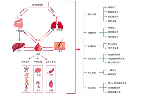
M/NPs能穿越组织屏障转移至血液, 随后到达全身各系统并在其组织和体液中不断积累, 最终产生毒性效应[77].M/NPs进入细胞后, 能引发多种毒性效应, 包括氧化应激、细胞毒性和基因毒性, 具体表现为细胞死亡、线粒体毒性、细胞膜损伤、DNA损伤和染色体畸变等, 进而在人体各系统中引发器官的局部炎症、菌群失调以及代谢紊乱等严重后果, 增加了相关疾病的发病风险, 甚至会引起肿瘤和癌症的发生(图 2).此外, 由于鼠类在一定程度上与人具有生理相关性, 常将鼠类用于研究M/NPs对有机体健康影响.然而动物模型实验存在成本高、周期长、不易操作和有违伦理等问题, 研究人员也尝试运用人体细胞和体外器官模型来研究M/NPs对人体健康风险.基于此, 下文综述了M/NPs对人体各个系统的潜在健康影响及其内在机制.
 素材来源于https://www.freepik.com图 2 摄入和吸入微/纳米塑料对人类健康造成的潜在风险Fig. 2 Potential risks of ingestion and inhalation of micro/nano-plastics to human health
3.1 消化系统
M/NPs首先影响消化系统.当食物依次通过口腔、食道、胃和肠道时, M/NPs会随之迁移, 其中一部分被排出体外, 另一部分则在体内积累, 以上M/NPs可能引发消化道炎症、屏障通透性增强和微生物失调等.Zhang等[72]推测PS-MPs可能通过破坏人类结肠上皮细胞线粒体电子传递链(ETC)以诱导线粒体去极化, 从而导致早期细胞凋亡.Luo等[78]研究发现摄入PS-MPs能够加重小鼠结肠炎症, 具体表现为结肠长度缩短、炎症加重、粘液分泌减少和结肠通透性增加.Fournier等[79]研究表明, M/NPs影响婴儿肠道菌群种类组成, 减少有益菌群丰度、增加有害菌群丰度, 并且菌群变化幅度受到M/NPs的尺寸、丰度和种类的影响.Tong等[80]研究发现, 幽门螺杆菌(H.pylori)在聚乙烯微塑料(PE-MPs)表面形成生物膜, 这加快了H.pylori在小鼠胃中的定居速度, 从而加剧胃部损伤和炎症.此外, 随着PS-MPs浓度和表面粗糙程度的增加, 人类结肠腺癌细胞(Caco-2细胞)的细胞膜完整性遭到破坏, 细胞存活率逐渐下降[81].小鼠摄入高浓度PE-MPs后出现明显的肠道炎症, 其与炎症相关跨膜受体蛋白和转录因子表达也随之升高[82].
肝脏作为人体内最大的代谢器官, 影响着脂肪的消化分解和脂溶性维生素的吸收.M/NPs可能对肝脏产生多种毒性, 包括氧化应激、细胞膜损伤、细胞纤维化、脂肪代谢混乱和能量代谢混乱[83~85].M/NPs通过诱导人类正常肝细胞(HL7702细胞)中细胞核DNA和mtDNA损伤, 以及激活cGAS/STING信号通路, 从而导致肝纤维化[83].斑马鱼经过M/NPs长期暴露, 其脂肪代谢混乱, 体重下降, 并且其脂肪酸代谢相关的基因表达水平出现降低趋势[86].此外, M/NPs的毒性受多种因素的影响, 包括M/NPs的物理化学特性、M/NPs与其他污染物之间的相互作用.Wang等[87]研究发现, 人工胃液能增强PS-MPs对肝细胞的毒害, 包括肝细胞的形态学改变、细胞膜损伤和氧化应激引起的细胞凋亡增加.Banerjee等[88]研究发现, 较小的微粒更容易被人肝癌细胞(HepG2细胞)内化, 相比羧基化或非功能化PS-M/NPs, 胺化PS-M/NPs对HepG2细胞毒性更大.此外, PS-MPs与双酚A(BPA)的联合暴露能干扰脂肪代谢、加剧肝毒性, 并且干扰与多种脂质代谢过程相关的基因等, 导致脂肪变性[89].
3.2 呼吸系统
肺是呼吸系统最重要的器官, 目前关于M/NPs对呼吸系统影响的研究主要是基于肺细胞开展的.M/NPs能够诱导肺细胞活性氧(ROS)增加和线粒体膜电位下降, 改变细胞正常结构, 从而提高急慢性呼吸系统疾病的发病风险.Zhang等[90]研究发现, 聚对苯二甲酸乙二醇酯纳米塑料(PET-NPs)诱导肺癌人类肺泡细胞(A549细胞)发生氧化应激和线粒体膜电位下降.Goodman等[91]研究发现, PS-MPs能诱发A549细胞形态改变, 使其细胞质突起增加和细胞接触丧失, PS-MPs通过影响人正常肺支气管上皮细胞(BEAS-2B细胞)间连接蛋白, 导致肺屏障功能障碍, 增加了慢性阻塞性肺疾病的风险[92].此外, M/NPs的尺寸影响其内化速度和肺部的微生物调节.相比20 nm和100 nm的PS-NPs, A549细胞对40 nm的PS-NPs的内化速度更快.这表明M/NPs可能存在内化速度相对较快的粒径范围[71].Zha等[93]研究发现, MPs与NPs均可诱发小鼠鼻腔和肺部微生物菌群失调, 但NPs比MPs对肺部微生物区系的影响更大.此外, 环境中M/NPs的丰度与暴露时间影响着肺部患病风险.例如, 合成纺织业和植绒业的工人长时间接触高丰度的M/NPs, 其患肺炎和慢性支气管炎风险远高于普通人[94].
3.3 生殖系统
不孕不育率的持续增长促使人们关注污染物对生殖系统的影响.M/NPs可能会引发细胞氧化应激、抗氧化活性降低和线粒体功能障碍, 进而引发生殖系统能量代谢失衡、激素紊乱和性器官损伤, 最终影响生殖能力下降, 造成不孕不育.M/NPs造成的雄性生殖毒性包括:睾丸质量下降、睾酮分泌减少、血睾屏障(BTB)遭受破坏和生精小管损伤, 从而导致生精功能障碍和精子畸形率上升, 以及精子活性和数量的下降[95~97].Jin等[98]研究证实, < 10 μm的MPs能进入睾丸细胞, 并在小鼠睾丸中积累.进入睾丸后, PS-MPs通过Hippo信号通路诱导幼鼠睾丸发育障碍[99], 通过氧化应激激活MAPK-Nrf2通路, 从而破坏大鼠BTB的完整性[97].此外, 常用的塑化剂邻苯二甲酸盐不仅会影响性激素分泌, 还能够增加PS-MPs导致的睾丸转录组改变, 诱导氧化应激[100].
M/NPs诱发的雌性生殖毒性包括卵巢毒性、发情期缩短和影响后代的发育与健康.其中, 卵巢毒性包括卵巢炎症、卵泡数量减少、卵巢细胞纤维化及细胞凋亡和颗粒细胞脱落[101~103].此外, 由于孕期和婴儿期是环境暴露的窗口期, 环境污染物容易影响胎儿的健康与发育.PS-NPs通过影响肌肉和脂质的代谢延缓小鼠胎儿生长, 导致其体重下降[104].PS-NPs能在小鼠间存在代际传递现象, 并能在子代大脑中积累, 导致子代神经干细胞功能、神经细胞组成变化和神经发育异常, 从而增加其神经发育缺陷和大脑功能障碍的风险[105].胎盘的主要功能是将营养物质从母体转运至胎儿, 提供胆固醇和类固醇激素以维持妊娠和胎儿发育.先前关于胎盘模型的研究表明, M/NPs的尺寸、表面特性、官能团和蛋白冠的不同组成能够影响胎盘对M/NPs的摄取和转运[66].人血白蛋白是提升M/NPs的转运的关键因素[106].此外, 一些塑料添加剂被视为内分泌干扰物, 需关注妊娠期暴露塑料添加剂对相关激素的干扰, 及其与胎盘功能相关基因之间存在的潜在相关性[107].
3.4 免疫系统
M/NPs对免疫系统的毒性主要包括免疫毒性、提高有害微生物和病原体的感染能力.其中, 巨噬细胞和淋巴细胞是M/NPs免疫毒性的主要对象.食品包装释放的N/MPs能够直接被小鼠巨噬细胞吸收和积累[108].PS-NPs进入人类THP-1巨噬细胞后, 能诱导ROS增加, 导致核损伤与线粒体膜电位下降, 从而降低细胞生存力.其中, 由不同结构和粒径组成的PS-NPs混合物能产生更高的细胞毒性, 甚至改变THP-1巨噬细胞形态[109].Çobanoğlu等[110]发现PE-MPs对人外周血淋巴细胞产生基因毒性, 增加其微核(MN)、核质桥(NPB)和核芽(NBUD)形成的频率.此外, M/NPs可能通过干扰免疫相关的基因和蛋白质影响免疫调节.PE-MPs暴露增加了小鼠血液内中性粒细胞数量及免疫球蛋白A(IgA)水平, 并改变脾脏内的淋巴细胞亚群结构[111].
受污染的M/NPs作为载体, 提高了病原体和有害微生物对人体的感染能力, 这对免疫系统健康造成了严重威胁.Wang等[112]研究发现, 大量甲型流感病毒(IAV)能够富集在PS-MPs上, 并通过内吞作用进入A549细胞中, 这促进了IAV对A594细胞的感染.Kampf等[113]研究发现, 冠状病毒能连续9 d在塑料表面保持传染性.紫外线辐射下老化的M/NPs对病毒有更强的吸附能力, 这能延长病毒存活期, 增加病毒传染性[114].此外, M/NPs能够在各种环境和生物之间传递抗生素抗性基因[115], 可能会增加有害微生物的抗生素耐药性.
3.5 心血管系统
有研究提示M/NPs可能会导致血液毒性和血管毒性, 并受M/NPs自身的物理化学特性影响.其中, 血液毒性包括M/NPs引发溶血、血栓和凝血.Barshtein等[116]研究表明, PS-NPs引发溶血受微粒的尺寸和丰度影响, 但人血白蛋白是最关键的影响因素, 可以阻止溶血发生.Wu等[58]研究推断, 环境微粒可能是血栓形成的核心, 初始血栓会持续吸引血液中的微粒, 以增大血栓的体积.Oslakovic等[117]研究发现, 胺基改性NPs与凝血相关因子Ⅶ和Ⅸ与结合能够减少凝血酶的形成.此外, M/NPs能够产生血管毒性.100 nm的NPs能在人脐静脉内皮细胞的细胞质中积累, 诱导自噬启动和自噬体形成, 这可能会引发细胞自噬过度和细胞坏死, 从而影响血管形成能力[68, 118].此外, M/NPs可能会导致心率异常和心脏功能受损.Chen等[119]研究发现, 在水环境暴露PS-M/NPs后, 海洋小鳉胚胎的血红蛋白和心脏发育相关基因表达受到影响.Pitt等[67]研究发现, PS-NPs在斑马鱼中出现代际传递现象, 并在子代中观察到心率过慢.Roshanzadeh等[120]研究发现, 胺化羧酸盐PS-NPs导致新生大鼠心肌细胞(NRVMs)收缩力降低, 影响方式随时间而变化.前期, 由细胞内Ca2+水平和电生理活动的降低导致NRVMs收缩力降低;晚期则由于线粒体膜电位和细胞代谢的下降导致NRVMs收缩力进一步下降.
4 展望
研究重点逐渐从最初探究M/NPs的环境分布状况与生态环境影响, 转移到评估M/NPs造成的人类健康的影响且研究成果颇多, 但目前仍存在许多知识空白:
(1)关于M/NPs人体暴露状况的研究, 大多针对环境介质进行检测分析, 关于综合暴露源头导致的暴露量评估极其匮乏.应结合不同暴露途径与暴露源, 以及地域、年龄、性别、职业和生活习惯等影响因素, 建立相关模型, 从而更为精确且全面地评估人体暴露量.
(2)以往研究的样本量较少, 实验数据受个体差异与检测方法影响较大.需要更多资源的支持以开展大规模的M/NPs人体富集状况的检测.
(3)小粒径和高浓度下M/NPs的富集和毒性有所增加.但毒理实验多选用粒径统一的PS微球作为暴露试剂.要考虑M/NPs在实际环境中的复杂性(如:尺寸、形状、聚合物组成、表面形态和风化程度等).特别是M/NPs作为载体, 与其他环境污染物和有害微生物病原体之间存在相互作用及联合毒性需要更多地关注.
(4)毒理研究集中于M/NPs高浓度急性暴露对人体健康的负面影响, 尚不清楚长期暴露的后果.然而实际环境状况中暴露浓度低且M/NPs的生物积累和降解程度会随着时间的推移而增加, 影响着M/NPs的生物毒性.因此, 基于低浓度和生命周期的M/NPs暴露对人体毒性的评估也是未来重要的研究方向.
5 结论
经口摄入是环境中的M/NPs最主要的暴露渠道, 食物、水和空气是最主要的环境介质, 应重视.M/NPs在人体中的积累较为普遍, 但丰度较低, 除结直肠腺癌肿瘤组织外, 约(702.68±504.26)n·g-1 M/NPs突破组织屏障后随着血液循环至全身各系统, 粒径较大的M/NPs虽然无法进入细胞, 但会损坏细胞膜, 较小的M/NPs能够进入细胞内并在细胞质中积累.M/NPs毒性受到其尺寸、形状、表面形态、聚合物类型、暴露途径、暴露时间和暴露浓度的影响, 此外M/NPs在体内降解中会释放有毒塑料添加剂, 并且M/NPs能将环境中其他有害污染物、微生物和病原体带入体内, 以上因素能够在细胞层面诱发氧化应激、细胞毒性、线粒体毒性和基因毒性等, 进而导致各系统炎症反应、屏障受损、代谢紊乱、内分泌紊乱以及菌群失调, 增加了各系统相关疾病的发病风险, 甚至会引起肿瘤和癌症的发生.然而, M/NPs对人体的毒害机制研究尚处于初步阶段, 动物和人对污染物的毒性反应有所差异, 细胞和器官模型不能模拟真实的人体内部状况, 其实验结果用于评估M/NPs对人体健康风险存在不足之处.
参考文献
[1] 刘微, 李宇欣, 荣飒爽, 等. 土壤中微塑料对陆生植物的毒性及其降解机制研究进展[J]. 环境科学, 2023, 44(11): 6267-6278.Liu W, Li Y X, Rong S S, et al. Research progress on toxicity of microplastics in soil to terrestrial plants and their degradation mechanism[J]. Environmental Science, 2023, 44(11): 6267-6278. [2] 张龙飞, 刘玉环, 阮榕生, 等. 微塑料的形成机制及其环境分布特征研究进展[J]. 环境科学, 2023, 44(8): 4728-4741.
Zhang L F, Liu Y H, Ruan R S, et al. Research progress on distribution characteristics and formation mechanisms of microplastics in the environment[J]. Environmental Science, 2023, 44(8): 4728-4741. [3] Wang J, Liu X H, Li Y, et al. Microplastics as contaminants in the soil environment: a mini-review[J]. Science of the Total Environment, 2019, 691: 848-857. DOI:10.1016/j.scitotenv.2019.07.209 [4] Cole M, Lindeque P, Halsband C, et al. Microplastics as contaminants in the marine environment: a review[J]. Marine Pollution Bulletin, 2011, 62(12): 2588-2597. DOI:10.1016/j.marpolbul.2011.09.025 [5] Zhang K, Hamidian A H, Tubić A, et al. Understanding plastic degradation and microplastic formation in the environment: a review[J]. Environmental Pollution, 2021, 274. DOI:10.1016/j.envpol.2021.116554 [6] Caldwell J, Taladriz-Blanco P, Lehner R, et al. The micro-, submicron-, and nanoplastic hunt: a review of detection methods for plastic particles[J]. Chemosphere, 2022, 293. DOI:10.1016/j.chemosphere.2022.133514 [7] 任欣伟, 唐景春, 于宸, 等. 土壤微塑料污染及生态效应研究进展[J]. 农业环境科学学报, 2018, 37(6): 1045-1058.
Ren X W, Tang J C, Yu C, et al. Advances in research on the ecological effects of microplastic pollution on soil ecosystems[J]. Journal of Agro-Environment Science, 2018, 37(6): 1045-1058. [8] Abbasi S, Alirezazadeh M, Razeghi N, et al. Microplastics captured by snowfall: a study in Northern Iran[J]. Science of the Total Environment, 2022, 822. DOI:10.1016/j.scitotenv.2022.153451 [9] Jiang C B, Yin L S, Li Z W, et al. Microplastic pollution in the rivers of the Tibet Plateau[J]. Environmental Pollution, 2019, 249: 91-98. DOI:10.1016/j.envpol.2019.03.022 [10] Crosta A, De Felice B, Antonioli D, et al. Microplastic contamination of supraglacial debris differs among glaciers with different anthropic pressures[J]. Science of the Total Environment, 2022, 851. DOI:10.1016/j.scitotenv.2022.158301 [11] Kanhai L D K, Gårdfeldt K, Lyashevska O, et al. Microplastics in sub-surface waters of the Arctic Central Basin[J]. Marine Pollution Bulletin, 2018, 130: 8-18. DOI:10.1016/j.marpolbul.2018.03.011 [12] Zhao J M, Ran W, Teng J, et al. Microplastic pollution in sediments from the Bohai Sea and the Yellow Sea, China[J]. Science of the Total Environment, 2018, 640⁃641: 637-645. [13] Beaumont N J, Aanesen M, Austen M C, et al. Global ecological, social and economic impacts of marine plastic[J]. Marine Pollution Bulletin, 2019, 142: 189-195. DOI:10.1016/j.marpolbul.2019.03.022 [14] Do A T N, Ha Y, Kwon J H. Leaching of microplastic-associated additives in aquatic environments: a critical review[J]. Environmental Pollution, 2022, 305. DOI:10.1016/j.envpol.2022.119258 [15] Gulizia A M, Patel K, Philippa B, et al. Understanding plasticiser leaching from polystyrene microplastics[J]. Science of the Total Environment, 2023, 857. DOI:10.1016/j.scitotenv.2022.159099 [16] Revel M, Châtel A, Mouneyrac C. Micro(nano)plastics: a threat to human health?[J]. Current Opinion in Environmental Science & Health, 2018, 1: 17-23. [17] Yu L Y, Li R L, Zhang Z, et al. Distribution, characteristics, and human exposure to microplastics in mangroves within the Guangdong-Hong Kong-Macao Greater Bay Area[J]. Marine Pollution Bulletin, 2022, 175. DOI:10.1016/j.marpolbul.2022.113395 [18] Nor N H M, Kooi M, Diepens N J, et al. Lifetime accumulation of microplastic in children and adults[J]. Environmental Science & Technology, 2021, 55(8): 5084-5096. [19] Cox K D, Covernton G A, Davies H L, et al. Human consumption of microplastics[J]. Environmental Science & Technology, 2019, 53(12): 7068-7074. [20] 张士春, 庞美霞, 赵洪雅, 等. 海产食品微纳塑料污染现状与危害[J]. 食品安全质量检测学报, 2019, 10(9): 2689-2696.
Zhang S C, Pang M X, Zhao H Y, et al. Situation and harm of micro-nano plastic pollution in seafood[J]. Journal of Food Safety and Quality, 2019, 10(9): 2689-2696. [21] Piyawardhana N, Weerathunga V, Chen H S, et al. Occurrence of microplastics in commercial marine dried fish in Asian countries[J]. Journal of Hazardous Materials, 2022, 423. DOI:10.1016/j.jhazmat.2021.127093 [22] Diaz-Basantes M F, Nacimba-Aguirre D, Conesa J A, et al. Presence of microplastics in commercial canned tuna[J]. Food Chemistry, 2022, 385. DOI:10.1016/j.foodchem.2022.132721 [23] Kılıç E, Yücel N. Microplastic occurrence in the gastrointestinal tract and gill of bioindicator fish species in the northeastern Mediterranean[J]. Marine Pollution Bulletin, 2022, 177. DOI:10.1016/j.marpolbul.2022.113556 [24] Debbarma N, Gurjar U R, Ramteke K K, et al. Abundance and characteristics of microplastics in gastrointestinal tracts and gills of croaker fish (Johnius dussumieri) from off Mumbai coastal waters of India[J]. Marine Pollution Bulletin, 2022, 176. DOI:10.1016/j.marpolbul.2022.113473 [25] Cho Y, Shim W J, Jang M, et al. Abundance and characteristics of microplastics in market bivalves from South Korea[J]. Environmental Pollution, 2019, 245: 1107-1116. DOI:10.1016/j.envpol.2018.11.091 [26] Muhib M I, Uddin M K, Rahman M M, et al. Occurrence of microplastics in tap and bottled water, and food packaging: a narrative review on current knowledge[J]. Science of the Total Environment, 2023, 865. DOI:10.1016/j.scitotenv.2022.161274 [27] Kosuth M, Mason S A, Wattenberg E V. Anthropogenic contamination of tap water, beer, and sea salt[J]. PLoS One, 2018, 13(4). DOI:10.1371/journal.pone.0194970 [28] Brei V A. How is a bottled water market created?[J]. WIREs Water, 2018, 5(1). DOI:10.1002/wat2.1220 [29] Kankanige D, Babel S. Smaller-sized micro-plastics (MPs) contamination in single-use PET-bottled water in Thailand[J]. Science of the Total Environment, 2020, 717. DOI:10.1016/j.scitotenv.2020.137232 [30] Yang D Q, Shi H H, Li L, et al. Microplastic pollution in table salts from China[J]. Environmental Science & Technology, 2015, 49(22): 13622-13627. [31] Kuttykattil A, Raju S, Vanka K S, et al. Consuming microplastics? Investigation of commercial salts as a source of microplastics (MPs) in diet[J]. Environmental Science and Pollution Research, 2023, 30(1): 930-942. DOI:10.1007/s11356-022-22101-0 [32] Iñiguez M E, Conesa J A, Fullana A. Microplastics in Spanish table salt[J]. Scientific Reports, 2017, 7(1). DOI:10.1038/s41598-017-09128-x [33] Lee H, Kunz A, Shim W J, et al. Microplastic contamination of table salts from Taiwan, including a global review[J]. Scientific Reports, 2019, 9(1). DOI:10.1038/s41598-019-46417-z [34] Conti G O, Ferrante M, Banni M, et al. Micro- and nano-plastics in edible fruit and vegetables. The first diet risks assessment for the general population[J]. Environmental Research, 2020, 187. DOI:10.1016/j.envres.2020.109677 [35] Dong Y M, Gao M L, Qiu W W, et al. Uptake of microplastics by carrots in presence of As (Ⅲ): combined toxic effects[J]. Journal of Hazardous Materials, 2021, 411. DOI:10.1016/j.jhazmat.2021.125055 [36] Liu Q R, Chen Z, Chen Y L, et al. Microplastics contamination in eggs: detection, occurrence and status[J]. Food Chemistry, 2022, 397. DOI:10.1016/j.foodchem.2022.133771 [37] Chen J H, Chen G H, Peng H Q, et al. Microplastic exposure induces muscle growth but reduces meat quality and muscle physiological function in chickens[J]. Science of the Total Environment, 2023, 882. DOI:10.1016/j.scitotenv.2023.163305 [38] Du F N, Cai H W, Zhang Q, et al. Microplastics in take-out food containers[J]. Journal of Hazardous Materials, 2020, 399. DOI:10.1016/j.jhazmat.2020.122969 [39] Zhou G Y, Wu Q D, Tang P, et al. How many microplastics do we ingest when using disposable drink cups?[J]. Journal of Hazardous Materials, 2023, 441. DOI:10.1016/j.jhazmat.2022.129982 [40] Li D Z, Shi Y H, Yang L M, et al. Microplastic release from the degradation of polypropylene feeding bottles during infant formula preparation[J]. Nature Food, 2020, 1(11): 746-754. DOI:10.1038/s43016-020-00171-y [41] Luo Y L, Gibson C T, Chuah C, et al. Raman imaging for the identification of Teflon microplastics and nanoplastics released from non-stick cookware[J]. Science of the Total Environment, 2022, 851. DOI:10.1016/j.scitotenv.2022.158293 [42] Yang X, Man Y B, Wong M H, et al. Environmental health impacts of microplastics exposure on structural organization levels in the human body[J]. Science of the Total Environment, 2022, 825. DOI:10.1016/j.scitotenv.2022.154025 [43] Sridharan S, Kumar M, Singh L, et al. Microplastics as an emerging source of particulate air pollution: a critical review[J]. Journal of Hazardous Materials, 2021, 418. DOI:10.1016/j.jhazmat.2021.126245 [44] Vianello A, Jensen R L, Liu L, et al. Simulating human exposure to indoor airborne microplastics using a Breathing Thermal Manikin[J]. Scientific Reports, 2019, 9(1). DOI:10.1038/s41598-019-45054-w [45] Yurtsever M. Tiny, shiny, and colorful microplastics: are regular glitters a significant source of microplastics?[J]. Marine Pollution Bulletin, 2019, 146: 678-682. DOI:10.1016/j.marpolbul.2019.07.009 [46] Saweres-Argüelles C, Ramírez-Novillo I, Vergara-Barberán M, et al. Skin absorption of inorganic nanoparticles and their toxicity: a review[J]. European Journal of Pharmaceutics and Biopharmaceutics, 2023, 182: 128-140. DOI:10.1016/j.ejpb.2022.12.010 [47] Vogt A, Combadiere B, Hadam S, et al. 40nm, but not 750 or 1, 500nm, nanoparticles enter epidermal CD1a+ cells after transcutaneous application on human skin[J]. Journal of Investigative Dermatology, 2006, 126(6): 1316-1322. DOI:10.1038/sj.jid.5700226 [48] Prow T W, Grice J E, Lin L L, et al. Nanoparticles and microparticles for skin drug delivery[J]. Advanced Drug Delivery Reviews, 2011, 63(6): 470-491. DOI:10.1016/j.addr.2011.01.012 [49] Ibrahim Y S, Anuar S T, Azmi A A, et al. Detection of microplastics in human colectomy specimens[J]. JGH Open, 2021, 5(1): 116-121. DOI:10.1002/jgh3.12457 [50] Cetin M, Miloglu F D, Baygutalp N K, et al. Higher number of microplastics in tumoral colon tissues from patients with colorectal adenocarcinoma[J]. Environmental Chemistry Letters, 2023, 21(2): 639-646. DOI:10.1007/s10311-022-01560-4 [51] Horvatits T, Tamminga M, Liu B B, et al. Microplastics detected in cirrhotic liver tissue[J]. eBioMedicine, 2022, 82. DOI:10.1016/j.ebiom.2022.104147 [52] Schwabl P, Köppel S, Königshofer P, et al. Detection of various microplastics in human stool[J]. Annals of Internal Medicine, 2019, 171(7): 453-457. DOI:10.7326/M19-0618 [53] Amato-Lourenço L F, Carvalho-Oliveira R, Júnior G R, et al. Presence of airborne microplastics in human lung tissue[J]. Journal of Hazardous Materials, 2021, 416. DOI:10.1016/j.jhazmat.2021.126124 [54] Jenner L C, Rotchell J M, Bennett R T, et al. Detection of microplastics in human lung tissue using μFTIR spectroscopy[J]. Science of the Total Environment, 2022, 831. DOI:10.1016/j.scitotenv.2022.154907 [55] Huang S M, Huang X X, Bi R, et al. Detection and analysis of microplastics in human sputum[J]. Environmental Science & Technology, 2022, 56(4): 2476-2486. [56] Jiang Y, Han J C, Na J, et al. Exposure to microplastics in the upper respiratory tract of indoor and outdoor workers[J]. Chemosphere, 2022, 307. DOI:10.1016/j.chemosphere.2022.136067 [57] Leslie H A, Van Velzen M J M, Brandsma S H, et al. Discovery and quantification of plastic particle pollution in human blood[J]. Environment International, 2022, 163. DOI:10.1016/j.envint.2022.107199 [58] Wu D, Feng Y D, Wang R, et al. Pigment microparticles and microplastics found in human thrombi based on Raman spectral evidence[J]. Journal of Advanced Research, 2022. DOI:10.1016/j.jare.2022.09.004 [59] Rotchell J M, Jenner L C, Chapman E, et al. Detection of microplastics in human saphenous vein tissue using μFTIR: a pilot study[J]. PLoS One, 2023, 18(2). DOI:10.1371/journal.pone.0280594 [60] Ragusa A, Svelato A, Santacroce C, et al. Plasticenta: first evidence of microplastics in human placenta[J]. Environment International, 2021, 146. DOI:10.1016/j.envint.2020.106274 [61] Zhu L, Zhu J Y, Zuo R, et al. Identification of microplastics in human placenta using laser direct infrared spectroscopy[J]. Science of the Total Environment, 2023, 856. DOI:10.1016/j.scitotenv.2022.159060 [62] Liu S J, Guo J L, Liu X Y, et al. Detection of various microplastics in placentas, meconium, infant feces, breastmilk and infant formula: a pilot prospective study[J]. Science of the Total Environment, 2023, 854. DOI:10.1016/j.scitotenv.2022.158699 [63] Ragusa A, Notarstefano V, Svelato A, et al. Raman microspectroscopy detection and characterisation of microplastics in human breastmilk[J]. Polymers, 2022, 14(13). DOI:10.3390/polym14132700 [64] Fournier S B, D'Errico J N, Adler D S, et al. Nanopolystyrene translocation and fetal deposition after acute lung exposure during late-stage pregnancy[J]. Particle and Fibre Toxicology, 2020, 17(1). DOI:10.1186/s12989-020-00385-9 [65] Medley E A, Spratlen M J, Yan B Z, et al. A systematic review of the placental translocation of micro- and nanoplastics[J]. Current Environmental Health Reports, 2023. DOI:10.1007/s40572-023-00391-x [66] Dusza H M, van Boxel J, van Duursen M B M, et al. Experimental human placental models for studying uptake, transport and toxicity of micro- and nanoplastics[J]. Science of the Total Environment, 2023, 860. DOI:10.1016/j.scitotenv.2022.160403 [67] Pitt J A, Trevisan R, Massarsky A, et al. Maternal transfer of nanoplastics to offspring in zebrafish (Danio rerio): a case study with nanopolystyrene[J]. Science of the Total Environment, 2018, 643: 324-334. DOI:10.1016/j.scitotenv.2018.06.186 [68] Lu Y Y, Li H Y, Ren H Y, et al. Size-dependent effects of polystyrene nanoplastics on autophagy response in human umbilical vein endothelial cells[J]. Journal of Hazardous Materials, 2022, 421. DOI:10.1016/j.jhazmat.2021.126770 [69] Ramsperger A F R M, Narayana V K B, Gross W, et al. Environmental exposure enhances the internalization of microplastic particles into cells[J]. Science Advances, 2020, 6(50). DOI:10.1126/sciadv.abd1211 [70] Lu Y Y, Cao M Y, Tian M P, et al. Internalization and cytotoxicity of polystyrene microplastics in human umbilical vein endothelial cells[J]. Journal of Applied Toxicology, 2023, 43(2): 262-271. DOI:10.1002/jat.4378 [71] Varela J A, Bexiga M G, Åberg C, et al. Quantifying size-dependent interactions between fluorescently labeled polystyrene nanoparticles and mammalian cells[J]. Journal of Nanobiotechnology, 2012, 10: do-39.. [72] Zhang Y T, Wang S L, Olga V, et al. The potential effects of microplastic pollution on human digestive tract cells[J]. Chemosphere, 2022, 291. DOI:10.1016/j.chemosphere.2021.132714 [73] Chen Q Q, Gao J N, Yu H R, et al. An emerging role of microplastics in the etiology of lung ground glass nodules[J]. Environmental Sciences Europe, 2022, 34(1). DOI:10.1186/s12302-022-00605-3 [74] Ouyang Z Z, Mao R F, Hu E D, et al. The indoor exposure of microplastics in different environments[J]. Gondwana Research, 2022, 108: 193-199. DOI:10.1016/j.gr.2021.10.023 [75] 艾鑫宇, 田洪钰, 岳鹏, 等. 大气环境中微塑料的检测技术研究进展[J]. 应用化工, 2023, 52(4): 1276-1282.
Ai X Y, Tian H Y, Yue P, et al. Research progress on detection technology of microplastics in the atmospheric environment[J]. Applied Chemical Industry, 2023, 52(4): 1276-1282. [76] 李臻阳, 杨书申, 徐亮, 等. 大气环境中微塑料污染及其分析技术的研究进展[J]. 环境化学, 2022, 41(4): 1114-1123.
Li Z Y, Yang S S, Xu L, et al. Progress on microplastics pollution and its analysis methods in the atmosphere[J]. Environmental Chemistry, 2022, 41(4): 1114-1123. [77] Tsou T Y, Lee S H, Kuo T H, et al. Distribution and toxicity of submicron plastic particles in mice[J]. Environmental Toxicology and Pharmacology, 2023, 97. DOI:10.1016/j.etap.2022.104038 [78] Luo T, Wang D, Zhao Y, et al. Polystyrene microplastics exacerbate experimental colitis in mice tightly associated with the occurrence of hepatic inflammation[J]. Science of the Total Environment, 2022, 844. DOI:10.1016/j.scitotenv.2022.156884 [79] Fournier E, Ratel J, Denis S, et al. Exposure to polyethylene microplastics alters immature gut microbiome in an infant in vitro gut model[J]. Journal of Hazardous Materials, 2023, 443. DOI:10.1016/j.jhazmat.2022.130383 [80] Tong X H, Li B Q, Li J, et al. Polyethylene microplastics cooperate with Helicobacter pylori to promote gastric injury and inflammation in mice[J]. Chemosphere, 2022, 288. DOI:10.1016/j.chemosphere.2021.132579 [81] Yu X Q, Lang M F, Huang D F, et al. Photo-transformation of microplastics and its toxicity to Caco-2 cells[J]. Science of the Total Environment, 2022, 806. DOI:10.1016/j.scitotenv.2021.150954 [82] Li B Q, Ding Y F, Cheng X, et al. Polyethylene microplastics affect the distribution of gut microbiota and inflammation development in mice[J]. Chemosphere, 2020, 244. DOI:10.1016/j.chemosphere.2019.125492 [83] Shen R, Yang K R, Cheng X, et al. Accumulation of polystyrene microplastics induces liver fibrosis by activating cGAS/STING pathway[J]. Environmental Pollution, 2022, 300. DOI:10.1016/j.envpol.2022.118986 [84] Goodman K E, Hua T, Sang Q X A. Effects of polystyrene microplastics on human kidney and liver cell morphology, cellular proliferation, and metabolism[J]. ACS Omega, 2022, 7(38): 34136-34153. DOI:10.1021/acsomega.2c03453 [85] Cheng W, Li X L, Zhou Y, et al. Polystyrene microplastics induce hepatotoxicity and disrupt lipid metabolism in the liver organoids[J]. Science of the Total Environment, 2022, 806. DOI:10.1016/j.scitotenv.2021.150328 [86] Zhao Y, Bao Z W, Wan Z Q, et al. Polystyrene microplastic exposure disturbs hepatic glycolipid metabolism at the physiological, biochemical, and transcriptomic levels in adult zebrafish[J]. Science of the Total Environment, 2020, 710. DOI:10.1016/j.scitotenv.2019.136279 [87] Wang L X, Wang Y X, Xu M, et al. Enhanced hepatic cytotoxicity of chemically transformed polystyrene microplastics by simulated gastric fluid[J]. Journal of Hazardous Materials, 2021, 410. DOI:10.1016/j.jhazmat.2020.124536 [88] Banerjee A, Billey L O, McGarvey A M, et al. Effects of polystyrene micro/nanoplastics on liver cells based on particle size, surface functionalization, concentration and exposure period[J]. Science of the Total Environment, 2022, 836. DOI:10.1016/j.scitotenv.2022.155621 [89] Cheng W, Zhou Y, Xie Y C, et al. Combined effect of polystyrene microplastics and bisphenol A on the human embryonic stem cells-derived liver organoids: the hepatotoxicity and lipid accumulation[J]. Science of the Total Environment, 2023, 854. DOI:10.1016/j.scitotenv.2022.158585 [90] Zhang H J, Zhang S Y, Duan Z H, et al. Pulmonary toxicology assessment of polyethylene terephthalate nanoplastic particles in vitro[J]. Environment International, 2022, 162. DOI:10.1016/j.envint.2022.107177 [91] Goodman K E, Hare J T, Khamis Z I, et al. Exposure of human lung cells to polystyrene microplastics significantly retards cell proliferation and triggers morphological changes[J]. Chemical Research in Toxicology, 2021, 34(4): 1069-1081. DOI:10.1021/acs.chemrestox.0c00486 [92] Dong C D, Chen C W, Chen Y C, et al. Polystyrene microplastic particles: in vitro pulmonary toxicity assessment[J]. Journal of Hazardous Materials, 2020, 385. DOI:10.1016/j.jhazmat.2019.121575 [93] Zha H, Xia J F, Li S J, et al. Airborne polystyrene microplastics and nanoplastics induce nasal and lung microbial dysbiosis in mice[J]. Chemosphere, 2023, 310. DOI:10.1016/j.chemosphere.2022.136764 [94] Prata J C. Airborne microplastics: consequences to human health?[J]. Environmental Pollution, 2018, 234: 115-126. DOI:10.1016/j.envpol.2017.11.043 [95] D'Angelo S, Meccariello R. Microplastics: a threat for male fertility[J]. International Journal of Environmental Research and Public Health, 2021, 18(5). DOI:10.3390/ijerph18052392 [96] Deng Y F, Chen H X, Huang Y C, et al. Polystyrene microplastics affect the reproductive performance of male mice and lipid homeostasis in their offspring[J]. Environmental Science & Technology Letters, 2022, 9(9): 752-757. [97] Li S D, Wang Q M, Yu H, et al. Polystyrene microplastics induce blood–testis barrier disruption regulated by the MAPK-Nrf2 signaling pathway in rats[J]. Environmental Science and Pollution Research, 2021, 28(35): 47921-47931. DOI:10.1007/s11356-021-13911-9 [98] Jin H B, Ma T, Sha X X, et al. Polystyrene microplastics induced male reproductive toxicity in mice[J]. Journal of Hazardous Materials, 2021, 401. DOI:10.1016/j.jhazmat.2020.123430 [99] Zhao T X, Shen L J, Ye X, et al. Prenatal and postnatal exposure to polystyrene microplastics induces testis developmental disorder and affects male fertility in mice[J]. Journal of Hazardous Materials, 2023, 445. DOI:10.1016/j.jhazmat.2022.130544 [100] Deng Y F, Yan Z H, Shen R Q, et al. Enhanced reproductive toxicities induced by phthalates contaminated microplastics in male mice (Mus musculus)[J]. Journal of Hazardous Materials, 2021, 406. DOI:10.1016/j.jhazmat.2020.124644 [101] Liu Z Q, Zhuan Q, Zhang L Y, et al. Polystyrene microplastics induced female reproductive toxicity in mice[J]. Journal of Hazardous Materials, 2022, 42. DOI:10.1016/j.jhazmat.2021.127629 [102] Huang J, Zou L P, Bao M, et al. Toxicity of polystyrene nanoparticles for mouse ovary and cultured human granulosa cells[J]. Ecotoxicology and Environmental Safety, 2023, 249. DOI:10.1016/j.ecoenv.2022.114371 [103] Hou J Y, Lei Z M, Cui L L, et al. Polystyrene microplastics lead to pyroptosis and apoptosis of ovarian granulosa cells via NLRP3/Caspase-1 signaling pathway in rats[J]. Ecotoxicology and Environmental Safety, 2021, 212. DOI:10.1016/j.ecoenv.2021.112012 [104] Chen G Q, Xiong S Y, Jing Q, et al. Maternal exposure to polystyrene nanoparticles retarded fetal growth and triggered metabolic disorders of placenta and fetus in mice[J]. Science of the Total Environment, 2023, 854. DOI:10.1016/j.scitotenv.2022.158666 [105] Jeong B, Baek J Y, Koo J, et al. Maternal exposure to polystyrene nanoplastics causes brain abnormalities in progeny[J]. Journal of Hazardous Materials, 2022, 426. DOI:10.1016/j.jhazmat.2021.127815 [106] Gruber M M, Hirschmugl B, Berger N, et al. Plasma proteins facilitates placental transfer of polystyrene particles[J]. Journal of Nanobiotechnology, 2020, 18(1). DOI:10.1186/s12951-020-00676-5 [107] Li J, Zeng X W, Liang X L, et al. Gestational exposure to plastic additives and associations with placental function-related genes[J]. Environmental Science & Technology Letters, 2023, 10(1): 86-92. [108] Deng J Y, Ibrahim M S, Tan L Y, et al. Microplastics released from food containers can suppress lysosomal activity in mouse macrophages[J]. Journal of Hazardous Materials, 2022, 435. DOI:10.1016/j.jhazmat.2022.128980 [109] Koner S, Florance I, Mukherjee A, et al. Cellular response of THP-1 macrophages to polystyrene microplastics exposure[J]. Toxicology, 2023, 483. DOI:10.1016/j.tox.2022.153385 [110] Çobanoğlu H, Belivermiş M, Sıkdokur E, et al. Genotoxic and cytotoxic effects of polyethylene microplastics on human peripheral blood lymphocytes[J]. Chemosphere, 2021, 272. DOI:10.1016/j.chemosphere.2021.129805 [111] Park E J, Han J S, Park E J, et al. Repeated-oral dose toxicity of polyethylene microplastics and the possible implications on reproduction and development of the next generation[J]. Toxicology Letters, 2020, 324: 75-85. DOI:10.1016/j.toxlet.2020.01.008 [112] Wang C, Wu W J, Pang Z F, et al. Polystyrene microplastics significantly facilitate influenza A virus infection of host cells[J]. Journal of Hazardous Materials, 2023, 446. DOI:10.1016/j.jhazmat.2022.130617 [113] Kampf G, Todt D, Pfaender S, et al. Persistence of coronaviruses on inanimate surfaces and their inactivation with biocidal agents[J]. Journal of Hospital Infection, 2020, 104(3): 246-251. DOI:10.1016/j.jhin.2020.01.022 [114] Lu J, Yu Z G, Ngiam L, et al. Microplastics as potential carriers of viruses could prolong virus survival and infectivity[J]. Water Research, 2022, 225. DOI:10.1016/j.watres.2022.119115 [115] Liu Y, Liu W Z, Yang X M, et al. Microplastics are a hotspot for antibiotic resistance genes: progress and perspective[J]. Science of the Total Environment, 2021, 773. DOI:10.1016/j.scitotenv.2021.145643 [116] Barshtein G, Arbell D, Yedgar S. Hemolytic effect of polymeric nanoparticles: role of albumin[J]. IEEE Transactions on NanoBioscience, 2011, 10(4): 259-261. DOI:10.1109/TNB.2011.2175745 [117] Oslakovic C, Cedervall T, Linse S, et al. Polystyrene nanoparticles affecting blood coagulation[J]. Nanomedicine: Nanotechnology, Biology and Medicine, 2012, 8(6): 981-986. DOI:10.1016/j.nano.2011.12.001 [118] Lee H S, Amarakoon D, Wei C i, et al. Adverse effect of polystyrene microplastics (PS-MPs) on tube formation and viability of human umbilical vein endothelial cells[J]. Food and Chemical Toxicology, 2021, 154. DOI:10.1016/j.fct.2021.112356 [119] Chen J C, Chen M Y, Fang C, et al. Microplastics negatively impact embryogenesis and modulate the immune response of the marine medaka Oryzias melastigma[J]. Marine Pollution Bulletin, 2020, 158. DOI:10.1016/j.marpolbul.2020.111349 [120] Roshanzadeh A, Oyunbaatar N E, Ganjbakhsh S E, et al. Exposure to nanoplastics impairs collective contractility of neonatal cardiomyocytes under electrical synchronization[J]. Biomaterials, 2021, 278. DOI:10.1016/j.biomaterials.2021.121175
相关知识
人体 100% 发现!厨房海绵每月脱落上万亿微塑料,需要担心吗?|对话专家
研究进展
环境污染与先天性心脏病的研究进展
全球环境与健康领域的研究热点,以及我国环境与健康研究面临的机遇与挑战
电磁辐射对人体健康影响的多组学研究进展
开屏帮你问专家|如何应对微塑料这个“隐形的健康杀手”?
肠道菌群利用膳食纤维及其与人体健康关系研究进展
瘦型非酒精性脂肪性肝病的特点及其研究进展
PM2.5污染与低出生体重发生风险关联的研究进展
妊娠期感染性疾病研究新进展(临床综述)
网址: 微塑料的人体富集及毒性机制研究进展 https://www.trfsz.com/newsview69275.html
推荐资讯
- 1从出汗看健康 出汗透露你的健 3873
- 2早上怎么喝水最健康? 3659
- 3习惯造就健康 影响健康的习惯 3303
- 4五大原因危害女性健康 如何保 3208
- 5连花清瘟、布洛芬等多款感冒药 2965
- 6男子喝水喉咙里像放了刀子一样 2465
- 710人混检核酸几天出结果?1 2229
- 8第二轮新冠疫情要来了?疾控中 2227
- 9转阴多久没有传染性?满足四个 2163
- 10补肾吃什么 补肾最佳食物推荐 2113


